Table of Contents
Introduction
Tuberculosis is a chronic, contagious, granulomatous disease characterized by the development of tubercle nodules followed by caseation and calcification, debility, and muscle wasting. The disease is transmissible between animal and human beings.
Etiology
As based on species of genus mycobacterium, affecting animals, humans, and birds, it is classified majorly into four types:
- Bovine type: Animals and human.
- Human type: Human and animals.
- Avian type: Fowls, pigs, horses and dogs.
- Volebacillus: Murrines species.
Different species of Mycobacterium affect different animals and human beings.
- Cattle: M.bovis and M.tuberculosis.
- Human – M.tuberculosis and M.bovis.
- Horses: M. bovis (majorly), M.tuberculosis and M.avium.
- Dogs: M. bovis, M. tuberculosis and M. avium.
- Pigs: Bovine, human and avian type.
- Birds: M.avium.
Epidemiology
Prevalence of infection
- Tuberculosis is prevalent worldwide both in animals and human beings.
- TB is endemic in Nepal both in animals and human beings.
Economic impact
- Apart from the mortality and productive loss.
- Disease is relatively benign in pigs.
- Discarding and condemning infected carcasses at slaughter.
Predisposing factors
- Overpopulation in small area.
- Purely intensive rearing.
- Inter current infection.
- Poor sanitation.
- Inadequate ventilation.
- vitamin A and C deficiency.
- Young age groups and malnourishment.
Source of infection
- Exhaled air, feces and infected meat.
- Nasal discharge, sputum and tracheal mucus.
- Contaminated inanimate and animate objects.
- Reproductive discharges.
- Feed, water and soil.
Transmission
- Nasal discharge and tracheal mucus.
- Ingestion: Communal use of feed and water troughs.
- Ingestion of tuberculosis infected animals milk and their products.
- Direct contact with infected animals.
- Inhalation of droplet nuclei from aerosol.
- Congenital infection by vertical transmission.
Host affected
- Cattle are the primary host highly susceptible for the infection.
- Pure bred and cross bred are highly affected as compared to zebu cattle.
- Sheep and goats more or less resistant to infection.
- Badgers, possums, pigs, dogs, primates, large cats and elephants other than cattle are affected and acts as maintenance host.
- Infected cattle are the main source for human infection.
Pathogenesis
Mechanism to develop infection
- The cells wall of the mycobacteria is constituted by 40% of the lipid which is of trehalos-6, 6’dimycolate and sulphur containing glycolipids.
- Virulent tubercle bacilli destroy the phagosome and causes failure of phagolysosomal fusion and so there is survival of mycobacteria within the phagocytic cells.
Primary Tubercle Complex or Ghon’s focus
- The mycobacteria which gain entry in the host by inhalation get lodged in the alveolar surface of the lung and its bronchial and thoracic lymphnodes.
- By ingestion the organism reaches pharyngeal and mesenteric lymphnodes get locked up there.
- Within the corresponding lymphnodes the mycobacteria undergoes multiplication and develop small tubercles. The focus of such infection in the lymphnodes given the condition “Primary complex of infection” or “Ghon’s focus”.
Post Primary dissemination and Stage of late generalization
- Through draining of lymphnodes, bacteria reaches the blood circulation and spread into other body cavities due to drop in immune response.
- As a result the bacteria overcome the killing effect by host immune mechanism and causes infiltration and necrosis of neutrophils.
- Together, the bacteria and the dead immune cells are surrounded by multi epitheloid cell layers which is called as Langhan’s giant cells or granulomas and further this is surrounded by lymphocytes and fibrous connective tissues.
Clinical signs
General form
- Affected animals become docile, lethargic but seems to be bright and alert.
- Cows with prominent miliary tubercle lesions are clinically normal.
- Progressive emaciation.
- Capricious appetite, fluctuating body temperature and rough / sleek hair coat.
- In spite of good appetite animal does not put up weight.
- All these general signs are pronounced following calving.
Respiratory form
- Silent or paroxysmal cough especially during early morning and chilled weather.
- Chronic cough due to bronchial pneumonia.
- Rapid respiration with dyspnoea revealed by auscultation and percussion of the chest due to enlargement of bronchial lymphnode and obstruction of air ways.
- Squeaking crackles are audible but noise audible over the caudal lobes.
- Enlargement of retropharyngeal lymphnode causes dysphagia and noisy breathing due to pharyngeal obstruction.
Reproductive form
- Metritis and inflammation of placenta leads to infertility, abortion and failure in conception.
- Nervous signs.
- In horse painful cervical osteomyelitis causes stiffness of neck and difficulty to eat.
Sheep and goats
- Bronchopneumonia and terminal dyspnea.
- Intestinal ulceration with diarrhea which is rare in cattle.
- Enlarged lymphnode of alimentary tract.
- Kids more progressive to early death.
Pigs
- TB lesion is cervical lymphnodes.
Horses
- Cervical vertebrae show osteomyelitis, and causes stiffness of neck.
Zoonosis
- It is an important zoonotic disease. There is an increase incidence with an increase spread from animal to human.
- M. bovis is responsible for 5-10% of human tuberculosis. Children getting infected via drinking of infected breast milk.
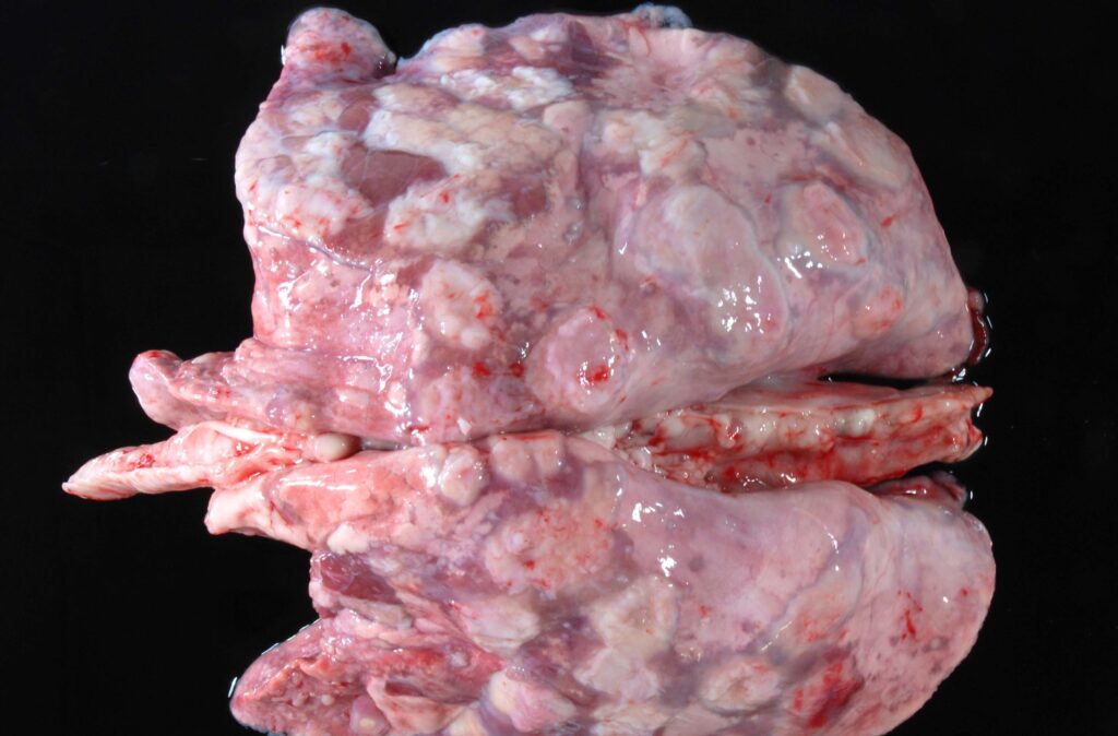
Necropsy findings
Cattle, sheep, goats
- Granulomatous lesion in bronchial, retropharyngeal AND mediastinal lymphnodes.
- Firm and enlarged pharyngeal lymphnode, swelling in dorsum of the pharynx.
- Miliary abscesses in lung.
- Pus is characteristic creamy and cheesy.
- TB nodule present in pleura and peritoneum.
- TB lesions are covered with fibrous capsule.
- Lesions in placenta with chronic purulent material.
- orchitis.
- Enlargement of supramammary lymphnode – TB lymphomatosis.
Source: cresa.cat

Source: Science Photo Library
Chronic lesions
- Discrete, nodular, thick yellow to orange caseous material followed by calcification covered by a thick fibrous capsule.
Pigs
- Enlarged miliary TB localized in tonsils, submaxillary, cervical, hepatic, bronchial, mediastinal, mesentric lymphnode with white caseous calcified material surrounded by a strong fibrous capsule.
Horses
- TB lesion in intestinal wall, mesenteric lymphnode and spleen.
- Cut surface of the LNs appears fleshy and resemble neoplastic tissue.
- Mineralisation and tissue necrosis noticed.
Diagnosis
- Based on clinical signs and necropsy findings.
- Palpation of supramammary lymphnode essential for suspected TB mastitis cases.
- Isolation and identification of organism by culture or acid fast staining.
- The following are the tests for detection of cell mediated immunity.
Single intradermal test
- PPD injection is given at caudal fold at base of the tail in sheep and goats / lateral center portion of the neck in cattle is highly sensitive.
- Initial skin thickness measured with a Vernier Caliper.
- DOSE: 0.1 ml herds with unknown status of sensitivity.
- 0.2 ml herds with known infected herds.
- Final skin thickness and inflammatory changes at 72 hours post injection should be taken.
- Increasing skin thickness more than 4mm is taken as positive.
Short thermal test
- Intradermal tuberculin 4ml injected s/c in the mid-neck of cattle.
- Initial rectal temperature 39 ° C (102 ° F) at the time of injection.
- If the temperature increase in 4, 6, 8hrs to above 40 ° C animal is a positive reactor.
Stormont test
- The first intradermal injection of 0.1 PPD at the middle of the neck followed by the second injection after 7 days at the same site.
- An increase in skin thickness of 5mm or more at 24 hrs after this second injection is test positive.
- Cattle with M. avium does not give a positive reaction but skin TB cases do.
- It is more accurate than SID.
Comparative test
- For testing the animals for TB, along the side of PPD tuberculin injection, 12cm apart, M.avium PPD should be given and the test result should be compared.
- Test results read after 72 hrs.
- This test is not used for initial screening.
- But can be used as a primary test when there is a high incidence of Avian TB and JD.
False-positive false-negative reaction
- Advance case of TB. Anergic animals are those with visible lesions of TB. But do not react to the intradermal DTH test.
- Early cases until 6 wks after injection.
- Cows that have calved within the preceding 6 weeks.
Gamma Interferon Assay
- This test measures the amount of gamma interferon released from the antigen-stimulated whole blood cells.
- Elisa for detection of antibodies.
- Molecular diagnosis by PCR.
- Sputum, feces, milk, blood, and tracheal secretions must be collected for the diagnosis.
Differential diagnosis
- Lung abscess.
- Pleurisy.
- Pericarditis.
- Traumatic reticulitis.
- Chronic contagious bovine pleuropneumonia.
- Upper respiratory tract infection.
- Actinobacillosis
- Bbovine leukosis.
- Lymphadenopathy.
- Mastitis.
Treatment
- There is no antibiotic treatment successful in control tuberculosis.
- Supportive treatment to enhance the immune response.
Prevention
- Test all animals over six months and above by tuberculin test.
- Positive reactors disposed of according to local legislation.
- Retest suspicious cases by comparative test.
- Retesting: If incidence is higher, retest 45-60 days after desensitization of intradermal injected animals.
- Annual testing of all cattle, quarantine,
- Slaughter of test positive herds
- Identification of individuals and wild life reservoir.
- BCG vaccination is available for calf hood vaccination. But vaccinated animals react positive to the skin test. Immunity is not strong and long lasting.
- Most probably cell mediated immunity is predominant.
Control
- Disinfection of utensils with 5% hot phenol and cresol.
- Feeding of calves with milk free from infection.
- Test and introduce new stock.